-
Die Universität
- Herzlich willkommen
- Das sind wir
- Medien & PR
-
Studium
- Allgemein
- Studienangebot
- Campusleben
-
Forschung
- Profil
- Infrastruktur
- Kooperationen
- Services
-
Karriere
- Arbeitgeberin Med Uni Graz
- Potenziale
- Arbeitsumfeld
- Offene Stellen
-
Diagnostik
- Patient*innen
- Zuweiser*innen
-
Gesundheitsthemen
- Gesundheitsinfrastruktur
Case of the Month
November 2024
Anal “tumour” in a 73-year-old female.
Diagnosis
Anal gland/duct cyst.
Comment
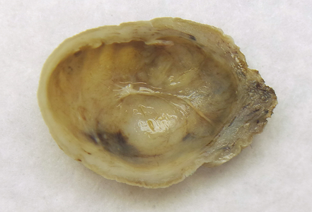
A 73-year-old woman underwent surgical excision of a polypoid lesion located in the anal region. The clinical description of the lesion on the request form was “giant marisca”. Gross examination showed a skin-covered specimen that contained a unilocular submucosal cystic lesion measuring about 2.8 cm in largest diameter. The cyst was filled with a slightly thick and brown-yellowish fluid, the cyst wall was smooth and thin incomplete septa were present (Panel A).
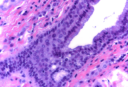
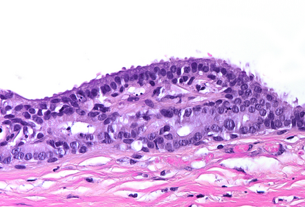
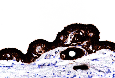
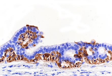
Histological examination disclosed a hybrid cyst lining epithelium, composed of a luminal layer of columnar cells with slightly basophilic cytoplasm and signs of mucin production, while slightly darker cuboidal cells without specific differentiation were present basally (Panel B-C). The cells exhibited mild nuclear irregularities, but no true dysplastic or malignant features were observed. The fibrotic cyst wall contained no skin adnexal structures, neural tissue, smooth muscle bundles or specific stroma. Immunohistochemical studies showed strong and diffuse reactivity for cytokeratin 7 of both epithelial layers (Panel D). In addition, the basal epithelial cell layer demonstrated immunoreactivity for cytokeratin 5/6 (Panel E) and p40 (Panel F). No immunohistochemical reactivity was observed for CDX2, CK20, PAX8, estrogen and progesterone receptors. Based on the morphological and immunohistochemical features, the diagnosis of a benign cyst lined by anal gland epithelium (“anal gland/duct cyst”) was made.
Anal gland cysts are uncommon cystic lesions of the (peri-)anal region that are assumed to represent retention cysts due to obstruction of the anal duct. These cysts are lined by columnar, squamous and/or transitional epithelium. The epithelium might show reactive change and mild cellular atypia, dysplasia or signs of invasive malignancy are usually absent.
Differential diagnosis of cystic lesions in the perianal region includes developmental cysts such as tailgut cysts, epidermoid and dermoid cysts, teratoma, anal duplication cysts or anterior meningocele. In addition, infectious and inflammatory lesions, cystic neoplasms and endometriosis cysts must be considered. Moreover, one must keep in mind that perianal cystic lesions can be complicated by secondary infection, fistulation or the development of dysplasia or neoplasia. Thus, complete excision and thorough sampling and examination is required for accurate histopathological diagnosis of perianal cystic lesions.
For further reading
- Brown IS, Sokolova A, Rosty C, and Graham RP. Cystic lesions of the retrorectal space. Histopathology. 2023; 82: 232-241.
- Seo GJ, Seo JH, Cho KJ, Cho HS. Anal Gland/Duct Cyst: A Case Report. Ann Coloproctol. 2020; 36: 204-6.
Presented by
Dr. Johanna Köhler, Linköping, Sweden and Dr. Cord Langner, Graz, Austria.


