-
Die Universität
- Herzlich willkommen
- Das sind wir
- Medien & PR
-
Studium
- Allgemein
- Studienangebot
- Campusleben
-
Forschung
- Profil
- Infrastruktur
- Kooperationen
- Services
-
Karriere
- Arbeitgeberin Med Uni Graz
- Potenziale
- Arbeitsumfeld
- Offene Stellen
-
Diagnostik
- Patient*innen
- Zuweiser*innen
-
Gesundheitsthemen
- Gesundheitsinfrastruktur

Case of the Month
November 2023
Whitish anal lesion in a 78-year-old female.
Diagnosis
Anal high-grade squamous intraepithelial lesion (HSIL).
Comment
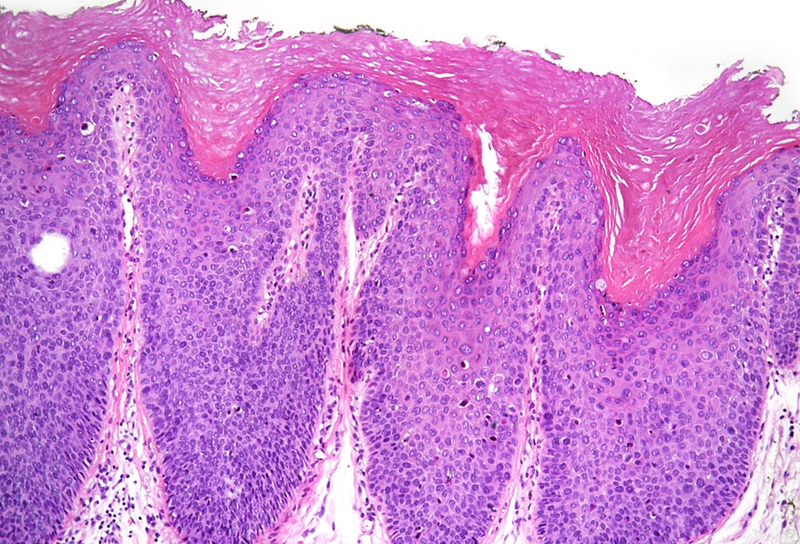
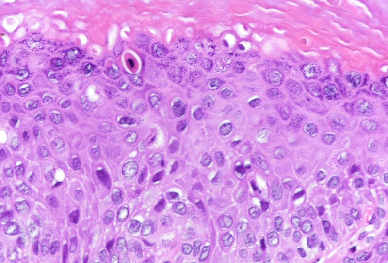
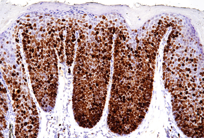
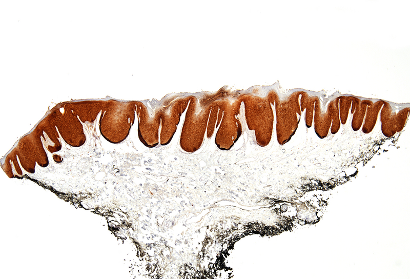
Microscopic examination revealed squamous dysplasia, affecting the full thickness of the epithelium (Panel A), with eventual koilocytes and increased apoptosis (Panel B). Mitotic figures were found throughout the epithelium, with marked increase in Ki-67 staining (Panel C). P16 immunohistochemistry reaction showed diffuse, block positivity which reached the excision margin (Panel D).
Anal high-grade squamous intraepithelial lesion (HSIL), also known as high-grade anal intraepithelial neoplasia (AIN III), is a premalignant condition, and a precursor of squamous cell carcinoma. It is associated with HPV infection, while further risk factors include high-risk sexual behavior, HIV infection, and immunocompromised states. Currently, there is no regular screening program anywhere regarding this disease, however, the identification of the affected population may serve later on as key. It has been suggested that genotyping HPV 16 could serve as the second step of the screening process, while other authors recommend the use of anal cytology, regardless of the high probability of contamination. Clinically, it usually remains asymptomatic, however, may present as anal bleeding, as well. Anoscopic findings include a plaque-like lesion, or a polypoid mass, therefore may be mistaken for hemorrhoid or rectal polyp. The primary treatment of anal HSIL consists of local excision or ablation, therefore, the incidence of anal squamous cell carcinoma may be significantly reduced. Topical therapy is not recommended.
Histologically, cytological and architectural atypia is visible is at least half of the thickness of the epithelium. Koilocytes may be present, as well. Mitotic figures may often be found in the upper half of the epithelium. Diffuse, block positivity of p16 may reflect HPV genome internalization, however, the definitive diagnostic tool of identification is polymerase chain reaction (PCR). Regarding differential diagnosis, several reactive epithelial changes, and metaplasias may mimic HSIL, especially on small biopsy samples. In such cases, p16 may play even more crucial role. Naturally, invasion has to be ruled out in each case.
For further reading
- Lyons KM, Butler SL. Anal intraepithelial neoplasia from a pathologists point of view. Clin Colon Rectal Surg. 2018;31:328-335.
- Ramirez R, Donohue K. Current monitoring and treatment for anal intraepithelial neoplasms. J Surg Oncol. 2023;127:1306-1309.
- Vyas M, Gonzalez RS. Anal intraepithelial neoplasia: a review of terminology, differential diagnoses, and patient management. Hum Pathol. 2023;132:56-64.
- Del Pino M, Matas I, Carrillo P, et al. Natural history of anal HPV infection in women treated for cervical intraepithelial neoplasia. Cancers (Basel). 2023;15:1147.
Presented by
Dr. Anita Sejben, Szeged, Hungary, Dr. Cord Langner, Graz, Austria.