-
Die Universität
- Herzlich willkommen
- Das sind wir
- Medien & PR
-
Studium
- Allgemein
- Studienangebot
- Campusleben
-
Forschung
- Profil
- Infrastruktur
- Kooperationen
- Services
-
Karriere
- Arbeitgeberin Med Uni Graz
- Potenziale
- Arbeitsumfeld
- Offene Stellen
-
Diagnostik
- Patient*innen
- Zuweiser*innen
-
Gesundheitsthemen
- Gesundheitsinfrastruktur

Case of the Month
July 2024
Biopsies from a 45-year-old male with endoscopic diagnosis of gastritis.

Diagnosis
Helicobacter heilmannii-associated gastritis.
Comment
A 45-year-old man with a history of chronic abdominal pain underwent gastroscopy. Endoscopically, erythematous antrum mucosa was observed, and gastric biopsies were taken.
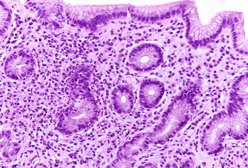
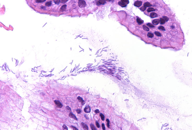
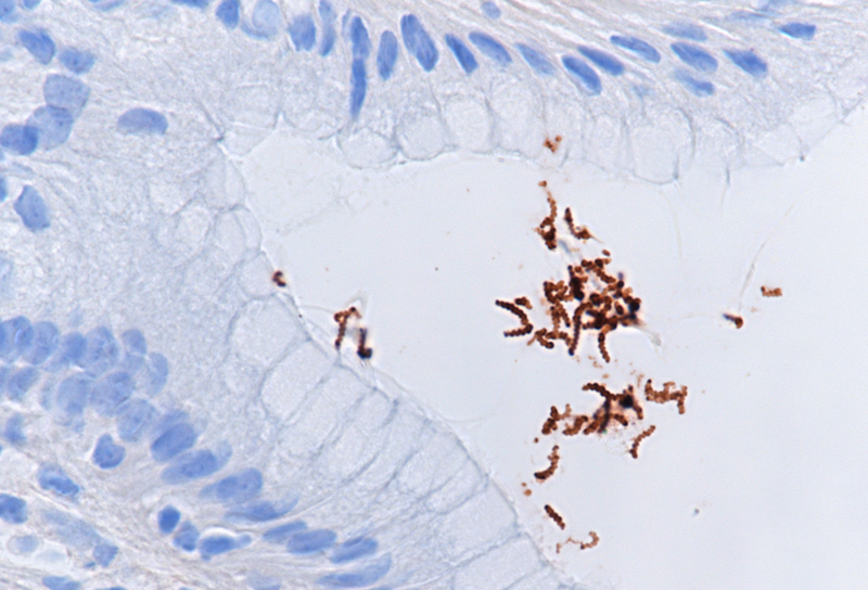
Histologic examination showed moderate chronic antrum gastritis with mild focal neutrophilic inflammation and small superficial mucosal erosions (Panel A). In the mucus layer close to the surface epithelium and in several of the foveolar pits, large, thin, corkscrew-shaped bacilli were found (Panel B) that could be highlighted with a H. pylori immunostain (Panels C-D). The characteristic morphology of the bacterial structures and their cross-reactivity with the anti-H. pylori antibody were consistent with Helicobacter heilmannii.
H. heilmannii-associated gastritis exhibits similar histological features to H. pylori-associated gastritis with chronic lymphoplasmacytic inflammation that is mainly located in the antrum. Occasionally, focal neutrophilic active inflammation, erosions and ulceration are observed. In comparison to H. pylori-associated gastritis, H. heilmannii-associated gastritis usually shows a lesser extent and milder degree of inflammation. Gastric H. heilmannii infection has been associated with intestinal metaplasia, gastric cancer and MALT-lymphoma.
Gastritis due to infection with Helicobacter heilmannii in humans is rare. The bacteria are more frequently found in cats, dogs, pigs and carnivorous mammals, and zoonotic transmission is assumed. Diagnosis is mainly based on histological examination of gastric biopsies, PCR and rapid urease testing can aid diagnosis. H. heilmannii-associated gastritis is treated with triple or quadruple eradication regimens similar to H. pylori treatment.
For further reading
- Heilmann KL, Borchard F. Gastritis due to spiral shaped bacteria other than Helicobacter pylori: clinical, histological, and ultrastructural findings. Gut 1991;32:137-140.
- Stolte, M., Kroher, G., Meining, A., Morgner, A., Bayerdörffer, E., & Bethke, B. (1997).
- A Comparison of Helicobacter pylori and H. heilmannii Gastritis: A Matched Control Study Involving 404 Patients. Scandinavian Journal of Gastroenterology, 32(1), 28–33.
- Joo M, Kwak JE, Chang SH, Kim H, Chi JG, Kim KA, Yang JH, Lee JS, Moon YS, Kim KM. Helicobacter heilmannii-associated gastritis: clinicopathologic findings and comparison with Helicobacter pylori-associated gastritis. J Korean Med Sci. 2007 Feb;22(1):63-9.
- Singhal, Anuradha V MD; Sepulveda, Antonia R MD, PhD. Helicobacter Heilmannii Gastritis: A Case Study With Review of Literature. The American Journal of Surgical Pathology 29(11):p 1537-1539, November 2005.
- Berry, Andrew et al. Not all Helicobacter are pylori. Clinical Gastroenterology and Hepatology, Volume 13, Issue 6, A23-A24. January 2015.
Presented by
Dr. Johanna Köhler, Linköping, Sweden and Dr. Cord Langner, Graz, Austria.