-
Die Universität
- Herzlich willkommen
- Das sind wir
- Medien & PR
-
Studium
- Allgemein
- Studienangebot
- Campusleben
-
Forschung
- Profil
- Infrastruktur
- Kooperationen
- Services
-
Karriere
- Arbeitgeberin Med Uni Graz
- Potenziale
- Arbeitsumfeld
- Offene Stellen
-
Diagnostik
- Patient*innen
- Zuweiser*innen
-
Gesundheitsthemen
- Gesundheitsinfrastruktur

Case of the Month
December 2023
Polypoid lesion at the anorectal junction in a 47 year-old male.
Diagnosis
Proctitis cystica profunda.
Comment
A 47-year-old male underwent haemorrhoidectomy. We received two irregular specimens, partially covered by anal and rectal mucosa, measuring up to 4.0:1.8:1.0 cm. Ectatic blood vessels were seen at the cut surface.
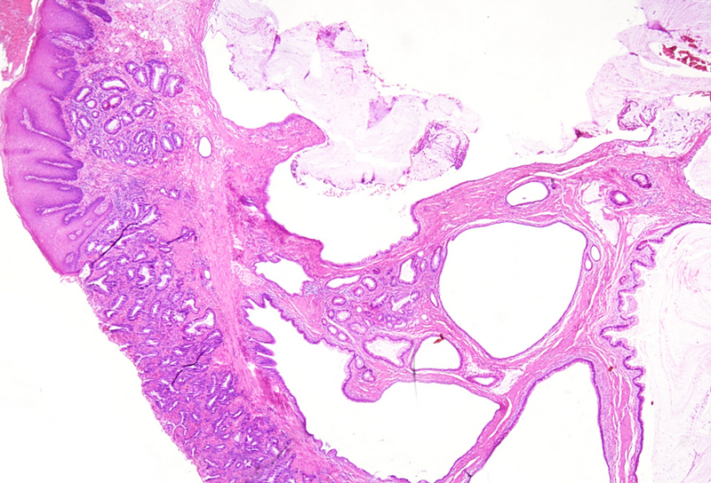
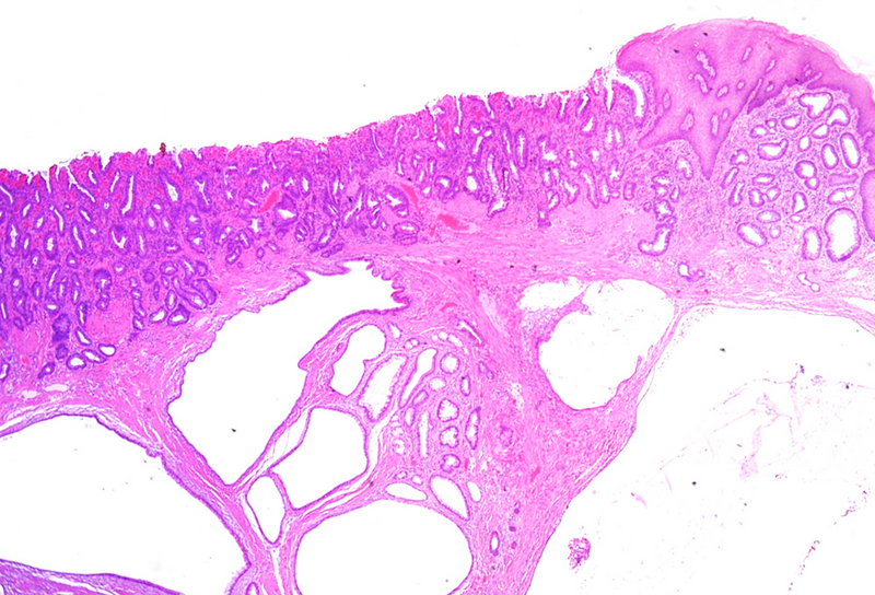
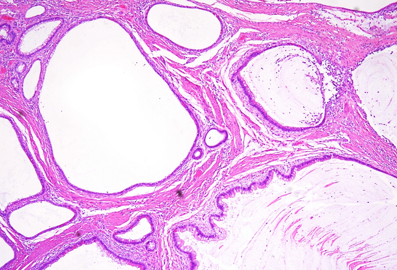
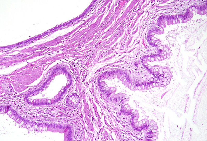
Microscopically, one specimen disclosed cystically dilated structures of variable size underneath regular colonic / anal mucosa (Panels A-B). The cysts are filled with mucin, and the epithelial lining is of intestinal type, showing reactive changes, yet no dysplasia in the presence of mixed unspecific inflammation (Panels C-D).
Colitis cystica profunda is a rare, non-neoplastic lesion, that may be observed in both colon and rectum. The aetiology remains enigmatic, however, it has been considered as a result of congenital, post-traumatic, and infectious states. At the anorectal junction, it has been related to solitary rectal ulcer syndrome and mucosal prolapse and may result from misplacement and entrapment of regenerative glands. Affected patients are usually in their third or fourth decade of life. Patients are mainly asymptomatic, or may present with diarrhoea or constipation, abdominal or anorectal pain, rectal bleeding, mucorrhoea, and tenesmus. While the clinical presentation may be similar to colorectal adenocarcinoma, biopsy sampling is advisory that can facilitate early diagnosis, therefore, avoid unnecessary surgical interactions, as well.
Histologically, it is characterized by the presence of either localised, segmental, or diffuse mucus-filled cysts of the deep submucosa and even muscularis propria. Size of the cysts may differ, depending on localisation and diffuse morphology, lesions can measure up to even 2 cm. The lining epithelium lacks atypia, with only minor nuclear irregularities in case of secondary inflammatory changes. The overlying mucosa is intact. Surrounding areas may contain fibrosis and hemosiderin laden macrophages. The cysts may rupture causing mucin dissemination into the surrounding tissue, potentially followed by degenerative calcification. Differential diagnosis mainly includes mucinous adenocarcinoma, which may be identified by nuclear dysplasia and/or desmoplastic response.
For further reading
- Papalampros A, Vailas MG, Sotiropoulou M, et al. Report of a case combining solitary Peutz-Jeghers polyp, colitis cystica profunda, and high-grade dysplasia of the epithelium of the colon. World J Surg Oncol. 2017;15:188.
- Jeruc J, Drobne D, Zidar N. Diffuse colitis cystica profunda in Crohn’s disease: A potential diagnostic pitfall. J Crohns Colitis. 2019;12:1362.
- Rumi N, Cilla S, De Ninno M, et al. Colitis cystica profunda of the rectum with adenomatous dysplastic features: Radiologic-pathologic correlation. Radiol Case Rep. 2019;14:740-745.
- Chen G, Jiang W, Yue M. Colitis cystica profunda of the rectum diagnosed by endoscopic submucosal dissection. Rev Esp Enferm Dig. 2023;115:91-92.
Presented by
Dr. Anita Sejben, Szeged, Hungary, Dr. Cord Langner, Graz, Austria.